800-333-5643  |  SALES  |  SERVICE  |  PAY INVOICE

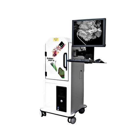
Kubtec XPERT 40

Easy to use, this mobile, self-contained, shielded cabinet X-ray system is designed to meet imaging requirements for surgically excised core tissue and stereotactic biopsies. The XPERT® 40 System eliminates scheduling conflicts in Mammography and reduces time in Surgery for intra-operative imaging of specimens. The XPERT® 40 System also has applications in Pathology for inspection of tissue specimens and wax blocks.

The XPERT® 40 System provides high resolution, high contrast, X-ray images, using either radiographic film, CR, or the DIGIVIEW™ line of digital detectors up to 8 x 8 inches (20 cm x 20 cm) in size. A small focal spot x-ray source and adjustable sample positioning provides the XPERT® 40 System with up to 5X magnification increasing visibility and detail of the smallest abnormalities. An x-ray source of up to 50kV and 1.0 mA provides easy penetration and eliminates the concerns of imaging dense surgical tissue in any specimen container.

The XPERT® 40 System has zero warm up time, AEC (automatic exposure control), auto-calibration, and the ability to send multiple images, with annotations, to PACS. Included with the standard DICOM compliant feature-rich software is the proprietary Artemis feature from Kubtec® that offers the unprecedented ability to view excised tissue with digital mammography quality. The touch screen monitor and mobile cart option make the XPERT® 40 System an easily maneuverable and user friendly unit offering the flexibility to meet the demands of every application.

When the demands are for versatility and high resolution imaging, look to the XPERT® 40 System from Kubtec, The Leader in Digital Specimen Radiography Systems.

With its complete shielding and other required radiation safety features, the XPERT® 40 System is in full compliance with US Federal and State requirements for radiation safety.

Brochure PDF

  • Largest detector size of all systems available in its class
  • Automatic calibration with zero warm up time
  • Ability to send multiple images to PACS with annotations
  • Up to 5X geometric magnification
  • Dedicated 3D imaging filter for enhanced visibility of micro-calcifications